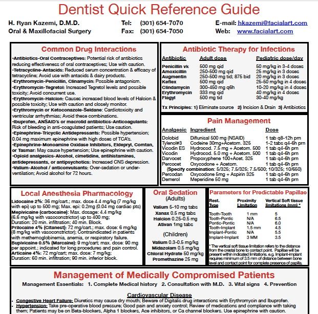
Dental Specialties Reference Guide
This article first appeared in the newsletter,. Not everything we do in dentistry is black and white and therein lies the conundrum, in that diagnosing and treating things that come our way is not as simple as we would like it to be. Oftentimes the chief complaint is difficult to pinpoint, and the pathology is asymptomatic or vague in appearance. Furthermore, lesions can be difficult to differentiate, and coming to a definitive diagnosis and subsequent treatment plan hinders our ability to serve our patients. With regard to that, it is no secret that internal and external resorption fall into this discussion.
- Dental Specialties Reference Guide
- Dental Specialties Reference Guide Indian Health Service
- Dental Specialties Reference Guide
In order to clear up what the gray area would be between these two lesions, these two basic questions need to be asked:. What are internal and external resorption, and what are the causes?. How do you differentiate between the two and come to a definitive diagnosis? The intent of this article is to submit succinct answers to those questions, and give practitioners a simple 101 guide that can be used as an adjunct to help offer predictable treatment and outcomes. The diagnosis of different types of internal and external resorption and specific treatment modalities are not discussed.
CIGNA REFERENCE GUIDE For physicians, hospitals. Specialty Care Physician (SCP). Dental, and Arizona. Introductory Guide for Commissioning Dental Specialties. 2 CLASSIFICATION. Cross Reference. 4 Why do NHS dental specialties need to change? Jump to Special Care Dentistry - Please note that the log book is compatible with both the DSCD and. Care Dentistry is available on request from the RCS Specialty. Includes case scenarios, structured reference, OSCE's and person.
ADDITIONAL READING What are internal and external resorption and the causes? The American Association of Endodontics defines resorption as, “a condition associated with either physiologic or a pathologic process resulting in a loss of dentin, cementum, and/or bone. (1) Vital tissue is necessary for either external or internal resorption to occur.” (2) By this definition, internal resorption is “a defect of the internal aspect of the root following necrosis of odontoblasts as a result of chronic inflammation and bacterial invasion of the pulp tissue.” (3) Contributing factors include caries, trauma, and restorative procedures. (3) External resorption is “resorption initiated in the periodontium and initially affecting the external surfaces of the tooth—may be further classified as surface, inflammatory, or replacement, or by location as cervical, lateral, or apical; may or may not invade the dental pulpal space.” (3) It may arise as a sequel of traumatic injury, orthodontic tooth movement, or chronic infection of the pulp or periodontal structures.” (4) ADDITIONAL READING How do you differentiate between internal and external resorption and come to a definitive diagnosis? Native Advertisement Being able to distinguish between internal and external resorption is often a challenge due to variations in tooth anatomy, unclear radiographs, and other underlying factors (i.e., restorations, etc.). An accurate definitive diagnosis will, therefore, depend on the practitioner’s understanding of what is “normal.” Radiographic interpretation is paramount, and Gartner, et al.
(2) produced a summary that assists in this differentiation (see table 1). Table 1: Radiographic appearance of internal and external resorption, early pulpal death, and dental caries In addition to this summary, Gartner et al. Discussed a radiographic aid called the mesial-buccal-distal (MBD) rule, which is used to determine the relative position of the roots.

(2) This technique is commonly used in endodontics. Take two radiographs.
First one: perpendicular to the tooth; second one: mesial to the perpendicular spot of the first one in the same horizontal plane. Closer objects: shift distally in relation to objects further from the source. With regard to internal and external resorption: If it’s external, “it will shift from its superimposed position over the canal system on the mesially angulated radiograph.” (2) If it’s internal, the “lesion will not shift no matter how severe an angle the radiograph was taken from, although the shape may change.” (2) In the event traditional radiographic resources have been exhausted or the diagnosis is still unclear, using 3-D imaging will allow for a complete assessment and subsequent diagnosis, as noted in Figures 1–4 below. This is a classic case of external resorption on the lingual of No.
19 (Images courtesy of Joseph A. Petrino, DDS, MS). Knowing what to look for when diagnosing internal and external resorption is paramount to what steps will subsequently be taken with regard to treatment, outcome and overall patient care.
Dental Specialties Reference Guide
Figure 1 Figure 2 Figure 3 Figure 4 Clinical examples I offer the four clinical examples below to help you distinguish between internal and external resorption. The outline of the canal in No. 24 is easily seen through the lesion, unaltered, and appearance is slightly ragged and irregular (figure 5). Diagnosis given to the patient at the time lesion was first noticed: internal resorption. As you can see, a root canal was completed, but no changes noted to the lesion. Patient continued to have discomfort post root canal therapy (figure 6).
Reassessment: external resorption confirmed by a 3-D scan. Tooth was recommended for removal and an implant was placed. Figure 5 Figure 6 Example No. This is another case of what could easily be misdiagnosed as internal resorption on tooth #No. If you look closely in Figure 7, the outline of the canal can be seen. In Figure 8, the lesion is advanced significantly (one-and-a-half years had gone by) the irregular borders and moth-eaten appearance is easily observed.

Definitive diagnosis: external resorption. Figure 7 Figure 8 Example No. (Courtesy of Christopher Shumway, DDS). Figures 9 and 10 show a discontinuation of the uniform outline of the pulpal chamber of tooth No. Furthermore, margins of the lesion are sharp and well defined. Diagnosis: internal resorption.
Figure 9 Figure 10 Example No. (Courtesy of Christopher Shumway, DDS). Figure 11 for tooth No. 25 shows a discontinuation of the uniform outline of the pulpal chamber; margins are also sharp and well defined. Diagnosis: internal resorption.
Dental Specialties Reference Guide Indian Health Service
Tooth was endodontically treated and has maintained a favorable outcome (figures 12 and 13). Figure 11 Figure 12 Figure 13 References 1.AAE Glossary of Endodontic Terms. American Association of Endodontists website. Accessed July 29, 2016.
Dental Specialties Reference Guide
2.Gartner AH, Mack T, Somerlott RG, Walsh LC. Differential diagnosis of internal and external root resorption. 3.Maria R, Mantri V, Koolwal S. Internal resorption: A review and case report. 4.Kuo T-C, Cheng Y-A, Lin C-P.
Clinical management of severe external root resorption. This article first appeared in the newsletter,. Simmons, DDS, is in private practice in Hamilton, Montana.
She is a graduate of Marquette University School of Dentistry. Simmons is a guest lecturer at the University of Montana in the Anatomy and Physiology Department. She is the editorial director of PennWell’s clinical dental specialties newsletter, DE’s Breakthrough Clinical with Stacey Simmons, DDS, and a contributing author for DentistryIQ, Perio-Implant Advisory, and Dental Economics. Simmons can be reached.
Nissan altima s 2015 manual. All motorcycle operating systems are explained in detail with easy-to-read text supported by high-quality illustrations. This manual is an invaluable resource for all students of motorcycle technology in general and Honda technology in particular. Each section begins with an outline of operational theory, continues with a detail of the various types of technology used by Honda over the years, and then follows up with troubleshooting and repair procedures.